Biofilm Workshop with Katja Bühler
Workshop Assignment
During the first Workshop on November 19th, we were asked by Dr. Katja Bühler to collect samples from different environments, surfaces or food to collect microbiological organisms. The goal was to practice working in a biological lab environment, sterile working and discussing the results in the second workshop. Another assignment as a practical example for biofilm formation was the production of vinegar via Wild Fermentation following a simple recipe with honey and water.
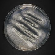
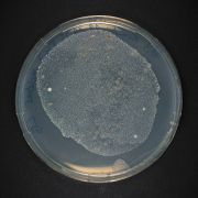
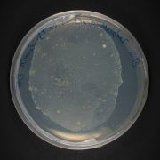
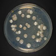
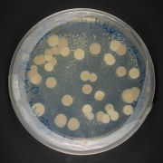
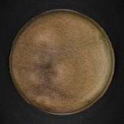
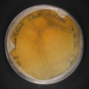
In the following, we present our beautiful petri dishes showing a variety of bacterial and fungal cultures collected from homemade fermented foods as Hibikus Kombucha, Kimchi, Milk and Water Kefir, as well as some environment samples from bio waste liquids and an isolated one year old water sample from the Kipperquelle (Weimar), which we collected 2021.
We photographed our petri dishes at home with a Fujifilm X-T3, a 7artisans Macro Lens 60mm f2.8 MK II and some macro extension rings on a stable inverted Tripod and some LED Lights from Godox.
| Security Information |
|---|
| Don't open Petri Dishes in sterile environments! Contamination of the lab should always be avoided! If you don't know what is inside, never open! Wear lab masks! |
Petri Dishes (Martin & Antje, 50 % each)
Martin was bringing in his experience/knowledge as a photographer and I was bringing in my experience/knowledge from 3 years working in the bio lab and with organisms and how to handle them.
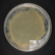
Bio Waste Liquid
Bio Waste Liquid
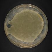
Hibiskus Kombucha

Hibiskus Kombucha
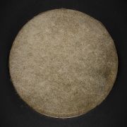
Kimchi Biofilm (Most propably Kahm Yeast)
Kimchi Biofilm (Most propably Kahm Yeast)
Kimchi Juice
Kimchi Juice
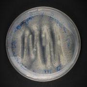
Kipperquelle Sample
Kipperquelle Sample
Sauerkraut Juice
Sauerkraut Juice


Macro Close Ups (Martin & Antje, 50 % each)
Martin was bringing in his experience/knowledge as a photographer and I was bringing in my experience/knowledge from 3 years working in the bio lab and with organisms and how to handle them.


(Antje) The pictures show nicely in how many different shapes and colors microbes can appear and as we later learned from the workshops with Zachery Denfeld what those shapes could tell you about the specimen and help roughly identify them for further research.
Microscopic Drawings (Antje)
We were also asked to make a drawing of what was seen under the microscope. This procedure has high educational value, since the microscopic image must be intensively observed. While drawing one needs to break down the structure that is seen to a much simpler one in order to achive an image that resamples the origin. It is also possible to represent specimens which cannot be photographed. The preparation of the drawing requires a great deal of time; short-lived specimens can therefore only be reproduced imperfectly. Last time I used this technique in high school. It was a challenge but also a great excercise which could help me in the future while observing something in nature and use it as an inspiration for my art works.
Drawing microscopic images has several advantages, including:
Documentation: Drawings provide a permanent record of the observations made under the microscope, allowing for easier comparison and study of the specimen over time.
Communication: Drawings can effectively communicate the details of microscopic observations to a wider audience, including other scientists and the general public.
Understanding: The act of drawing a specimen can help the observer better understand and analyze its features and characteristics.
Clarity: Drawings can reveal details that may not be immediately apparent through simple observation under the microscope, leading to a deeper understanding of the specimen.
Overall, drawing microscopic images has played a crucial role in the advancement of the field of microscopy and our understanding of the microscopic world.
Formation of Biofilm in wild fermented vinegar (Antje)
Vinegar Making assignment to do at home with a simple recipe using just water and honey and a bit of organic apple vinegar as a starter. Hopefully a mother of vinegar would form.
Was does the term "wild" fermentation refer to?
Wild fermentation is a process of fermenting food or drink where naturally occurring yeast and bacteria are used, without adding specific strains. It is a less controlled and less predictable method, as the outcome depends on the microorganisms present in the environment.
Controlled fermentation, on the other hand, involves adding a specific strain of yeast or bacteria to the food or drink to be fermented. This method allows for more consistent results and greater control over the flavor and other qualities of the final product. It is typically used in commercial food and beverage production to ensure consistent quality and flavor. Whereas commercial foods never have the variety of microbes that made at home ferments provide due to regulatories.
After one week some white wine was added to speed up the process. Normally during wild fermentation of vinegar, naturally occurring yeasts and acetic acid bacteria (AAB) convert alcohol into acetic acid. The process starts with the yeast converting sugar into alcohol through fermentation. Then, AAB consume the alcohol and produce acetic acid, which gives vinegar its characteristic sour flavor.
A small mother of vinegar formed (biofilm). This mother can be used for making also other kinds of vinegar. It normally speeds up the process naturally.
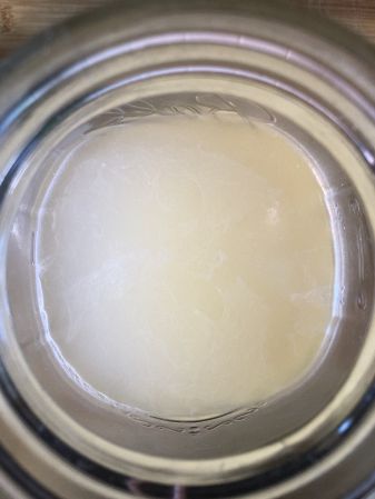
mother of vinegar formed on top
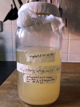
jar with wild fermented apple vinegar


Note The method mentioned above is a very simple one. There are more sophisticated processes with which one reaches a more advanced product richer in flavour. Normally one should let time do the work and don't speed up processes. Vinegar making needs time and once its "finished" even longer time to process in a dark well tempered place. For example NOMA, the renowned restaurant in Copenhagen, Denmark, is known for its innovative and unique approach to cooking and fermentation. They development methods making vinegar by combining traditional approaches with Innovation.
Endophyte Club with Zachery Denfeld
Workshop Assignment
During the Workshop on from December 2nd to December 3rd, we were asked by Zachery Denfeld to collect samples from different foods of the local supermarket REWE and prepare those samples for the studies of endophytes growing within our samples. We were introduced into the lab routine of preparing plant samples for endophyte identification.
In the following, we present or beautiful petri dishes showing a variety of bacterial and funghoid cultures collected from Paprika, Chives, Lamb's Lettuce, Coconut Fibre, Red Radish, Radish and Banana.
We photographed our petri dishes at home with a Fujifilm X-T3, a 7artisans Macro Lens 60mm f2.8 MK II and some macro extension rings on a stable inverted Tripod and some LED Lights from Godox.
| Security Information |
|---|
| Don't open Petri Dishes in sterile environments! If you don't know what is inside, never open or replate! Wear a mask. Contamination of the lab should always be avoided! |
Petri Dishes (Martin & Antje, 50 % each)
Martin was bringing in his experience/knowledge as a photographer and I was bringing in my experience/knowledge from 3 years working in the bio lab and with organisms and how to handle them.


Macro Close Ups (Martin & Antje, 50 % each)
Martin was bringing in his experience/knowledge as a photographer and I was bringing in my experience/knowledge from 3 years working in the bio lab and with organisms and how to handle them.


Identification (Antje)
Another goal of the workshop was to try to roughly identifiy what was growing on the petri dishes. Zachery was providing us with a table that gave a very basic idea of what could possibly be found or what to look for. More or less a differentiation between Form, Elevation and Margin could be made. We tried to identifiy ours. The most obvious differentiation one could achieve with the eyes or by observing with the microscope would be to identify wether the cultures are bacterial origin or fungal.
Fungal colonies and bacterial colonies in culture on a petri dish can differ in terms of their appearance, texture, and growth patterns. Under a microscope, fungal colonies typically appear as fluffy, white or off-white mats with a texture that is soft and sometimes cottony. Bacterial colonies, on the other hand, often appear as smooth, round or irregularly shaped, and opaque. Bacterial colonies may also have a more dense appearance compared to fungal colonies.
In terms of growth patterns, fungal colonies tend to spread outwards in a radial fashion and can form complex branching structures. Bacterial colonies, on the other hand, tend to grow in a circular fashion and do normally not form the same types of branching structures as fungi.
These differences can be used to distinguish between fungal and bacterial colonies under the microscope. However, it's important to note that some species of bacteria can form complex, three-dimensional structures, while some fungal species may form colonies that are relatively smooth and circular, making a definitive identification based solely on appearance challenging in some cases.
Conclusion
So what was again clear after the workshop there are several methods that can be used to definitively identify a specimen as either fungal or bacterial in nature.
Some of these include:
Microscopy: Examination of the microscopic structure of the colony can provide information about the nature of the organism. This may include observation of cell shape, size, and arrangement, as well as the presence of structures such as hyphae (in fungi) or flagella (in some bacteria).
Staining techniques: Different staining techniques can be used to highlight specific features of the cells and can aid in the identification of the organism. For example, Gram staining is commonly used to differentiate between Gram-positive and Gram-negative bacteria.
Molecular methods: DNA-based techniques, such as polymerase chain reaction (PCR) and sequencing, can be used to identify the organism to the species level. This may involve amplifying and sequencing specific genetic markers, such as the ribosomal DNA, that are characteristic of a particular group of organisms.
Culture characteristics: The way in which the colony grows on a petri dish can also provide information about the nature of the organism. This may include the rate of growth, the formation of aerial structures, and the ability to produce pigments or other metabolic products.
It is important to note that a combination of these methods may be required for a definitive identification, as some organisms may be difficult to classify based on a single criterion. In addition, some organisms may be misclassified due to contamination or other factors, so it is important to confirm the identity of the specimen through multiple lines of evidence.
But anyhow. As and Artist it is a good start to be curious and be inspired by the different shapes and colors. And to use this as a creative source for other projects.
Mapping my surrounding area (OBERWEIMAR/EHRINGSDORF) for endophytic cultures (Antje)
Artistic Study
I would like to map the area arround where I live in Oberweimar/Ehringsdorf for Endophytes and see how the shapes and colors could be a source of inspiration for the work with fabric, yarn and textile materials. I would like to explore the themes of these unseen biological systems and find a connection that could enhance my work with fabrics and change my point of view, also in terms of organic and reusable material or biomaterials:
Patterns The intricate patterns created by endophytic growth within plant tissues can inspire to create similar patterns, perhaps using weaving, knotting, or embroidery techniques.
Colony formation: Endophytes can form distinct colonies on the agar surface, each with a characteristic shape, size, and color. Zone of inhibition: Endophytes can produce compounds that inhibit the growth of other microorganisms, creating a clear zone around the endophyte colony. Spread patterns: Endophytes can spread out from the initial point of inoculation, creating a circular or radial pattern of growth. Mixed culture patterns: Endophytes can co-exist and interact with other microorganisms in the culture, creating complex and dynamic patterns of growth.
Color Endophytic organisms can add color to their host plants, one can use these colors as inspiration perhaps by dyeing or printing fabrics with natural pigments.
Texture The textures created by endophytic growth can be used as inspiration for the creation of similar textures through the use of different fabrics, fibers, or stitching techniques.
This artistic study could be the basis for greater Project with fabric.