No edit summary |
No edit summary |
||
| (6 intermediate revisions by the same user not shown) | |||
| Line 1: | Line 1: | ||
[[File:HEaderAmelia.jpg|1000px]] | |||
This Semester I want to explore and learn about Physarum Polycephalum in Order to Work with it in future artistic works. Through different experiments and approaches I want to find a sophisticated project Idea. | |||
'''Clothing and Physarum Polycephalum''' | |||
''' | |||
''04.06.2022'' Observing the growth in Petridish | ''04.06.2022'' Observing the growth in Petridish | ||
<gallery> | |||
File:ppinpetri.jpeg | |||
</gallery> | |||
''05.06.2022'' | ''05.06.2022'' | ||
| Line 38: | Line 22: | ||
'''(B)''' Placing the Organism in the Center of the Tshirt. It is not wet or feed with additional food or water. The Agar moisture spreads out on the fabric. I hope this will create a differet sturcture in growth than from '''A'''. Fabric: Satin | '''(B)''' Placing the Organism in the Center of the Tshirt. It is not wet or feed with additional food or water. The Agar moisture spreads out on the fabric. I hope this will create a differet sturcture in growth than from '''A'''. Fabric: Satin | ||
<gallery> | |||
File:Tshiört.jpeg | |||
File:Tisirt.jpeg | |||
File:Tshiiiii.jpeg | |||
</gallery> | |||
''06.06.2022'' Growth of '''A''' and '''B''' | ''06.06.2022'' Growth of '''A''' and '''B''' | ||
<gallery> | |||
File:666b222.jpeg | |||
File:6662222.jpeg | |||
</gallery> | |||
''08.06.2022'' Final day | ''08.06.2022'' Final day | ||
| Line 50: | Line 40: | ||
I really like the result of this one. | I really like the result of this one. | ||
<gallery> | |||
File:likki.jpeg | |||
File:pplieky.jpeg | |||
File:ppthstlke.jpeg | |||
</gallery> | |||
'''A''' | |||
As seen, for the moment to take the pictures, a part of the organism has already started going black. And the thickness of the rest has been reduced. It started dying. | |||
<gallery> | |||
File:deadppp.jpeg | |||
File:deadppp.jpeg | |||
File:deadpp.jpeg | |||
</gallery> | |||
'''Scoby''' | |||
For this Experiment I was following this guide. After prepearing and mixing I took it home to observe the bacterias growth. | |||
https://www.uni-weimar.de/kunst-und-gestaltung/wiki/GMU:Home_Made_Bioelectronics/Other_resources | |||
<gallery> | |||
File:Bacteriaa.jpeg | |||
</gallery> | |||
The 1 and 2 show the same behaviour. Small dots grow all over the petri dish just mass/coverage is decreased. In the dished 3 , 4 and 5 a different growth structure is visible. In these a | |||
serried growth from the edge to the middle is visible. Additionally small dots appear in 3 there are few dots than in 1:1000 and no rhytms in the growth from the side. Comparing 4 and 5 to the rest: Both show structure and rhythm in the growth from the side, the rest doesn´t. This might occur due to the mold growing inside which grew on accident. Comparing 4 and 5 to another the difference in the thickness and mass of the dots and patterns, rhythmsis visible. 4 has more dots than 3 and 5 which goes againt the "theory" the dots decreasing. | |||
Also in the reference dish 6 a small space is covered in bacteria which should not have happend. | |||
I can not explain why the behaviour, patterns changes nor why the intesity changes from 4 and 5. | |||
Revision as of 12:30, 21 June 2022
This Semester I want to explore and learn about Physarum Polycephalum in Order to Work with it in future artistic works. Through different experiments and approaches I want to find a sophisticated project Idea.
Clothing and Physarum Polycephalum
04.06.2022 Observing the growth in Petridish
05.06.2022
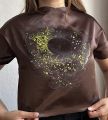
(A)Carefully placing the Organism on the T-shirt with a cardboard beneath. Then wetting and feeding with oats just around the aga cover. In order to control grow a cover is put over. Fabric: Cotton
(B) Placing the Organism in the Center of the Tshirt. It is not wet or feed with additional food or water. The Agar moisture spreads out on the fabric. I hope this will create a differet sturcture in growth than from A. Fabric: Satin
06.06.2022 Growth of A and B
08.06.2022 Final day
B I really like the result of this one.
A
As seen, for the moment to take the pictures, a part of the organism has already started going black. And the thickness of the rest has been reduced. It started dying.
Scoby
For this Experiment I was following this guide. After prepearing and mixing I took it home to observe the bacterias growth.
https://www.uni-weimar.de/kunst-und-gestaltung/wiki/GMU:Home_Made_Bioelectronics/Other_resources
The 1 and 2 show the same behaviour. Small dots grow all over the petri dish just mass/coverage is decreased. In the dished 3 , 4 and 5 a different growth structure is visible. In these a serried growth from the edge to the middle is visible. Additionally small dots appear in 3 there are few dots than in 1:1000 and no rhytms in the growth from the side. Comparing 4 and 5 to the rest: Both show structure and rhythm in the growth from the side, the rest doesn´t. This might occur due to the mold growing inside which grew on accident. Comparing 4 and 5 to another the difference in the thickness and mass of the dots and patterns, rhythmsis visible. 4 has more dots than 3 and 5 which goes againt the "theory" the dots decreasing. Also in the reference dish 6 a small space is covered in bacteria which should not have happend.
I can not explain why the behaviour, patterns changes nor why the intesity changes from 4 and 5.
References
- https://class.textile-academy.org/2020/loes.bogers/files/recipes/bacterialdye/
- https://www.lampoonmagazine.com/article/2021/08/06/vienna-textile-lab-bacterial-dyes-karin-fleck/
- https://www.scientificamerican.com/article/the-environments-new-clothes-biodegradable-textiles-grown-from-live-organisms/
- https://www.uni-weimar.de/kunst-und-gestaltung/wiki/GMU:Different_Worlds/Anna_Wissmueller
Artists
- https://annadumitriu.co.uk/
- https://www.realitydisfunction.org/?page_id=182
- https://www.google.com/search?rlz=1C1CHBF_deDE984DE984&source=univ&tbm=isch&q=decalcomanie+max+ernst&fir=JEWjxFlDBH3bvM%252ClXvJO1Yxdkt9OM%252C_%253B4l-9SSiGcDMGoM%252CTrYvdNcSjDY75M%252C_&usg=AI4_-kQer15CgRlQzDt5OwKB9GQyfMWeew&sa=X&ved=2ahUKEwjuw9qXsZb4AhXgSvEDHZC2ASsQ7Al6BAgaEAI&biw=1368&bih=761&dpr=2
Old Concept/Experiment with Martin Howse sensor


